Fetal echocardiography (echo) is a test that shows pictures of a baby’s heart before birth. The pictures are formed using mild sound waves. This is called ultrasound.
The test checks for problems in the baby’s heart structure, function, or rhythm. Finding these heart problems before birth means that they can be managed early. This may also help in planning for what to do after birth. Many heart problems can be found with fetal echo. But some can’t be seen until after the baby is born.
The test is painless. Nothing is put into your body. In rare cases, the ultrasound may need to be done in the vagina with a special wand. This may be the case very early in a pregnancy.
Why might I need a fetal echo?
The test may be done when you are at least 12 weeks pregnant. Your healthcare provider may advise this test if you:
-
Had a pregnancy ultrasound that showed a possible heart problem
-
Had problems found by other tests, such as amniocentesis or chorionic villus sampling (these check for genetic diseases and chromosomal problems)
-
Have a family history of congenital heart disease
-
Are taking medicines that may affect your baby’s development
-
Have a family history of genetic diseases linked with heart defects and disease
-
Have diabetes or other conditions
How should I get ready for a fetal echo?
You don't need to do much to get ready. Wear comfortable clothes. Follow any instructions you are given.
What happens during a fetal echo?
The test takes about 45 minutes to 2 hours. During the test:
-
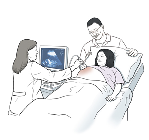
You lie on an exam table with your belly uncovered.
-
Clear, non-greasy gel is applied to the skin on your belly.
-
A hand-held probe (transducer) is moved across your belly.
-
Sound waves from the transducer go to a computer. Pictures of your baby’s heart are seen on a screen.
What happens after a fetal echo?
-
You can return to your normal routine and diet.
-
Your healthcare provider may talk to you about the early results right after the test. You will get the final results when the images have been fully studied.
There are no known risks with a fetal echo.
Featured in
Online Medical Reviewer: Callie Tayrien RN MSN
Online Medical Reviewer: Scott Aydin MD
Online Medical Reviewer: Stacey Wojcik MBA BSN RN
Date Last Reviewed: 09/01/2022
© 2000-2026 The StayWell Company, LLC. All rights reserved. This information is not intended as a substitute for professional medical care. Always follow your healthcare professional's instructions.